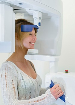
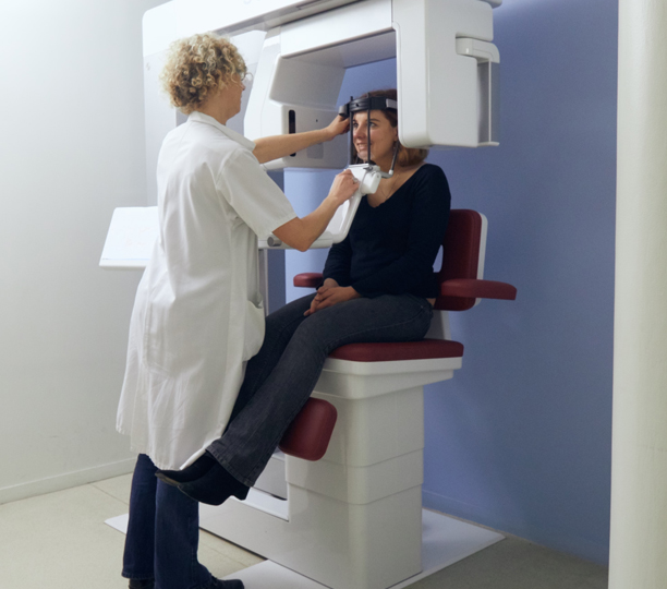
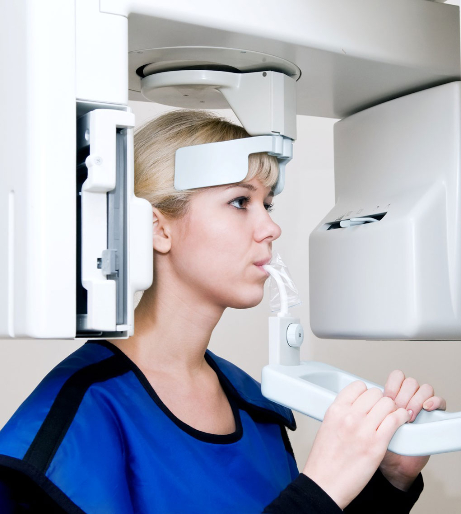
Scanner Cone Beam Sinus
3d Cone Beam Imaging Brooks Dental P C

Cone Beam Ct Scanner Lawrenceville Georgia Northeast Endodontics Inc

Cone Beam Ct Scan Loveland Fort Collins Co

What Is A Dental Cone Beam Ct Midtown Dental

Dr Ganesh At Santa Monica Esthetic Dentistry Cone Beam Ct

3d Cone Beam Scan Technology Seabreeze Dental Group

This article will be providing evidence on the diagnostic and treatment planning applications of cbct in sinus imaging mainly.
Scanner cone beam sinus.
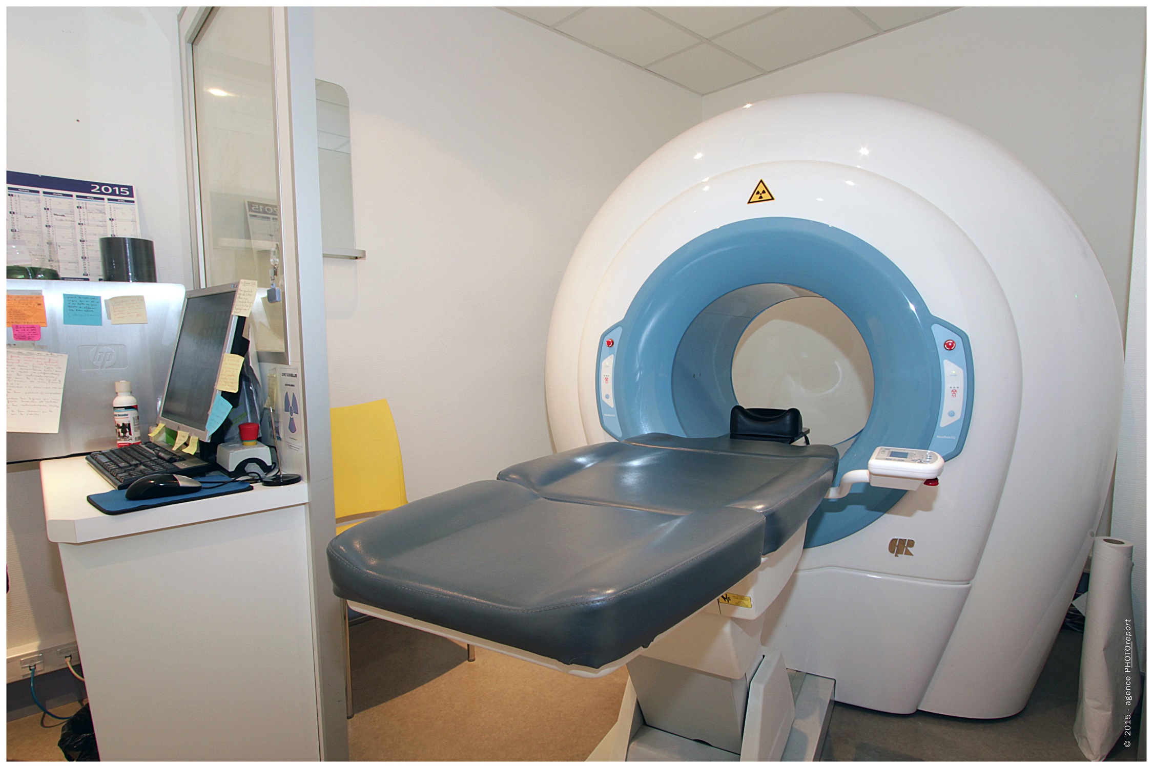
Cone Beam Presentation Centre Imagerie Point Medical Imagerie Point Medical
Cone Beam Scanner Technology Dr Asinmaz
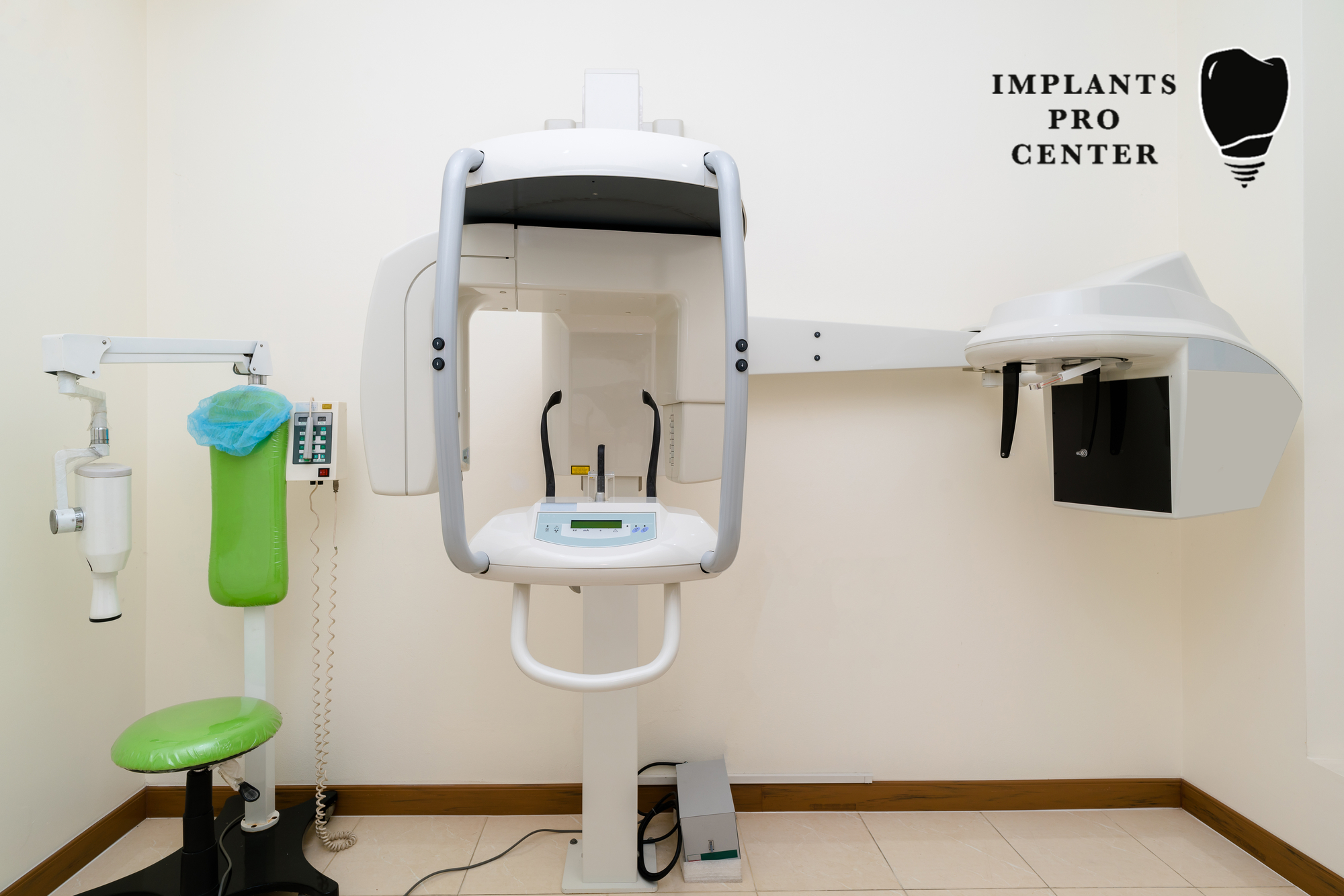
3d Cone Beam Advantages Applications Procedure Implants Pro Center C

Iluma Ultra Cone Beam Ct Scanner Midjersey Smiles

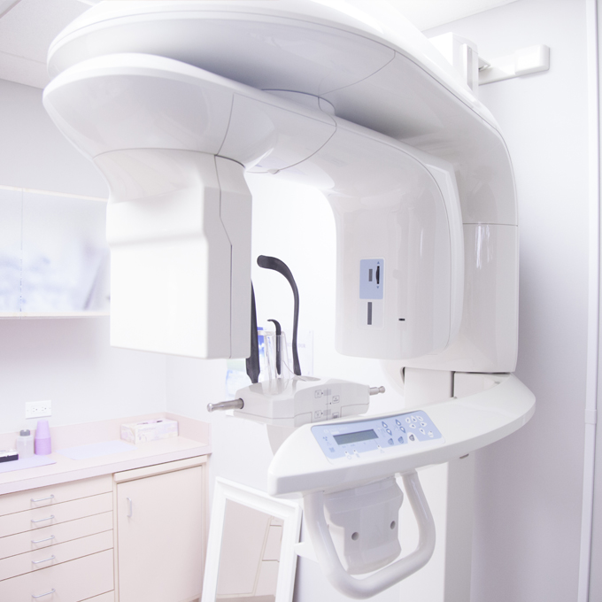
Cone Beam Scanner At Kk Dental Treatment With The Latest Technology
Cone Beam Iris Radiologie

Cone Beam 3d Scan Technology Glen Mills Pa Brandywine Periodontics

Le Scanner Cone Beam

Cone Beam Radiologie Et Imagerie Medicale Bartholdi

Cone Beam Ct Scanner Corvallis Or Valley Endodontics Root Canal Implant Dentistry

Cone Beam Scanner Dietrich Kelso Orthodontics Lakeland Winter Haven Brandon

Cone Beam Ct Scanner Cutting Edge Scanning Technology Dsny

3d Imaging Springboro Oh I Cat System Cone Beam Ct Technology
3d Cone Beam Ct Scanning

Cone Beam Ct Technology Children Adult Dentistry Fort Myers

3d Imaging Henrico Va Cone Beam Ct
Excel Dental Care Stream 3d Cone Beam Scanner

Sterling Dentist Our New 3d Dental Cone Beam Ct Scanner Is Here
Https Encrypted Tbn0 Gstatic Com Images Q Tbn 3aand9gcq421f0lv Ioxezoz I8p9traekkqna4p Kpxy5nqqzbyx3wje Usqp Cau

Perfect Smile Dental 3d Cone Beam Imaging

3d Imaging Mcmurray Pa Cone Beam Ct Washington County Pa

Sinus Elevation Livingston Periodontal Implant Associates

Galileos 3d Sunrise Dental Center

3d Imaging Jacksonville Fl Precision Diagnostics Dental Ct Scan
Source : pinterest.com
